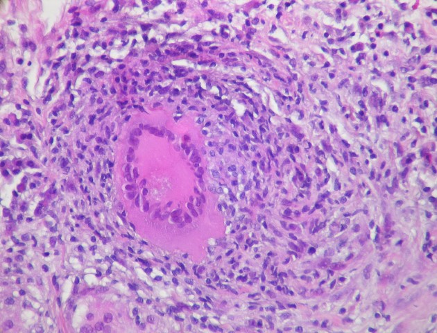
robably the most tested histological feature on the boards is the granuloma. If you can recognize the granuloma on a histology image, you can learn a lot about the disease process they are describing in the vignette and narrow your differential diagnosis significantly. As a caveat, I am not a pathologist, and a pathologist may cringe at my description below; but for the boards this description will help you find the granulomas almost every time.
Here are the features of a granuloma you want to look out for on a histology image:
- Circular shape:
- Blue cells:
- Pink cells:
- Multinucleated giant cells:
- The granuloma will probably be in the middle of the image:
- Information in the vignette:
There will be a bunch of cells in a conglomerate within a tissue that are seeming to form a circle. There will not be a distinct boundary so you may have to squint a little to convince yourself that there is indeed a circle of cells in the middle of the histology image.
The borders of the large circle of cells are made of mostly small blue cells with scant cytoplasm which represent mostly lymphocytes. If the entire circular mass of cells is made of small blue cells then you are probably dealing with a lymphoid follicle, so be aware of the other two cell types required below.
In the middle of the circle of cells there is usually a mass of pink cells that represent histiocytes.
These are the most important players, and the most defining characteristic of the granuloma. Look for a large pink cell with many nuclei. If you see one of these in a circle of cells on a histology slide, you can hang your hat on the fact that you are dealing with a granuloma.
The boards examiners are not trying to fool you and they don’t expect you to be a pathologist. If you see a circular blob of cells in the middle of the page with some small blue cells and you happen to spot a couple larger pink cells with several nuclei, then you have a granuloma.
Sometimes there will be information in the vignette to drive you to be looking for a granuloma in the first place. For example, if the vignette describes an African American patient with shortness of breath and hypercalcemia you should probably be looking for granulomas in your histology image.
Hooray! You’ve identified a granuloma, now what? Well, now you have an instant differential diagnosis, and a decently limited one at that. Here are a few of the most commonly tested granulomatous diseases:
- Granulomatosis with polyangiitis
- Tuberculosis
- Sarcoidosis
- Foreign body
- Fungal infections
- Crohn disease


